Portfolio
About
Team: who we are
Services: what we do
Process: how we work
Clients: who we work with
Testimonials: what they say
Certified Medical Illustrator
News
Contact
A gallery of our work
All Projects
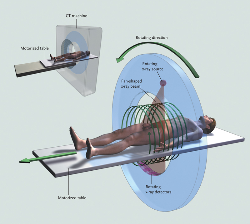
Basics of CT
»
Next
Previous
»